

Practice-changing refinements of lung cancer staging
The 8th edition of the TNM classification has recently come into effect. Compared to the 7th edition published in 2009 [1], several important adjustments have been made to lung cancer staging with the aim of improving prognostication and research [2]. “Research is of particular significance in the context of smaller tumours that can be treated with a variety of therapeutic options,” said Ramón Rami-Porta, MD, PhD, Department of Thoracic Surgery, Hospital Universitari Mútua Terrassa, Terrassa, Barcelona, Spain [3]. The new edition is based on a very large number of patients, which supports its robustness [4].
Differentiation of small tumours
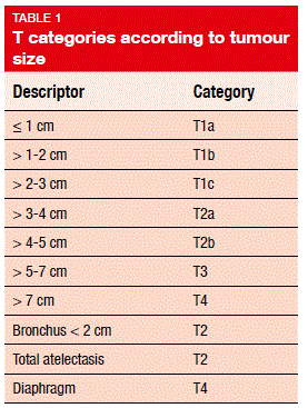
Changes with regard to the T descriptors account for the diversity of small tumours in the lungs, which can be identified routinely in the clinic using modern computed tomography techniques. “Tumours that had not been defined until 2011 have received names,” Dr. Rami-Porta stressed. For instance, this applies to adenocarcinoma in situ (Tis [AIS]) and minimally invasive adenocarcinoma (T1mi). In the past, the term of ‘Tis’ denoted only squamous-cell carcinoma. “It is important to make a difference here, because a patient can have both,” Dr. Rami-Porta explained. The smallest solid tumour, which corresponds to the new T1a category, has a maximum diameter of 1 cm (Table 1).
Awareness of smaller tumours will be raised due to the new categorisation. Also, these tumours can be used to study new treatment options, such as stereotactic radiotherapy, radiofrequency ablation, microwaves, or their combinations. Minimal resection is of interest here as well, as is the study of tumour biology in general. “Some tumours, like adenocarcinoma in situ, will not be resected right away, but instead be observed, allowing for the assessment of factors like growth and density.”
N and M changes
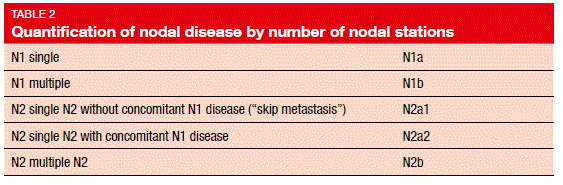
For quantification of lymph node involvement, nodal stations can be used, as well as the nodal zones that were defined by the 7thedition. “There are five possibilities to quantify nodal disease based on stations,” Dr. Rami-Porta said (Table 2). These correspond to four prognostic groups, as patients with N1b and N2a1 have the same prognosis. The numbers of involved lymph nodes, nodal zones, and nodal stations are of importance. This is also true for the lymph node ratio, which is calculated as the ratio of involved and removed nodes. “All of this can only be assessed during pathological staging,” Dr. Rami- Porta indicated.
In the area of M descriptors, the new classification states that the number of M1 lesions matters more than the location. Extrathoracic metastasis has been refined, with separation of single from multiple extrathoracic metastases in either one organ or several organs. “The sub-classification of extrathoracic metastases will contribute to a homogeneous definition of oligometastatic disease and oligoprogression, which are quite loosely defined nowadays,” Dr. Rami-Porta pointed out.
Recommendations on measurement and staining
Another innovation relates to the assessment of tumour size in partially solid tumours. For computed tomography assessment, only the solid part is of interest, as it appears to be equivalent to the invasive component. The pathologist determines the T category according to the size of the invasive component only, regardless of the size of the whole tumour, including any portions that show lepidic growth. “This innovation will change our practice,” Dr. Rami-Porta emphasised. “We were used to measuring the size of the entire tumour.”
Moreover, it is recommended to use elastic stains to identify invasion of the visceral pleura, which was shown to be an important prognostic factor. Dr. Rami-Porta noted that staging can change according to the extent of pleural invasion. “If this is not clear enough according to haematoxylin and eosin stains, it is important to use elastic stains.” When lung cancer with multiple primary lesions is present, TNM staging is assigned to each tumour. In multiple adenocarcinomas with ground-glass opacity/ lepidic features, the highest T category is used, and the number of tumours is given in parentheses.
“The innovations in the 8th edition of TNM staging for lung cancer will increase our capacity to refine prognosis, improve tumour stratification in future trials, prompt future research, and facilitate homogeneous tumour classification, as well as collection of prospective data,” Dr. Rami-Porta concluded.
REFERENCES
- Sobin LH et al., TNM classification of malignant tumours, 7th Edition. November 2009, Wiley-Blackwell, ISBN: 978-1-4443-3241-4
- Goldstraw P et al., The IASLC Lung Cancer Staging Project: Proposals for revision of the TNM stage groupings in the forthcoming (eighth) edition of the TNM classification for lung cancer. J Thorac Oncol 2016; 11(1): 39-51
- Rami-Porta R, Lung cancer staging – changing the clinical practice, WCLC 2016, PL03.02
- Rami-Porta R et al., The IASLC lung cancer staging project: the new database to inform the eighth edition of the TNM classification of lung cancer. J Thorac Oncol 2014; 9: 1618-1624